|
![]() |
|||
|
||||
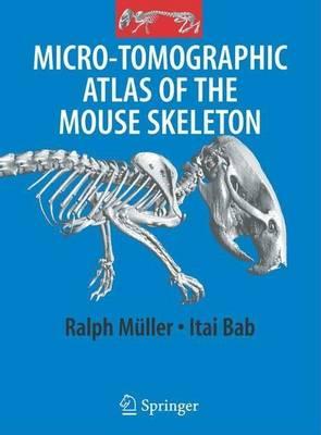
OverviewThe Micro-Tomographic Atlas of the Mouse Skeleton provides a unique systematic description of all calcified components of the mouse. It includes about 200 high resolution, two- and three dimensional m CT images of the exterior and interiors of all bones and joints. In addition, the spatial relationship of bones within complex skeletal units (e.g., skull, thorax, pelvis, extremities) is also described. The images are accompanied by detailed explanatory text, thus highlighting special features and newly reported structures. With mice becoming a standard laboratory animal and the rapidly increasing use of m CT technology as a key analytical tool in skeletal research, the Atlas fulfils an emerging need for a comprehensive reference to assist both trained and in-training researchers. Full Product DetailsAuthor: Itai Bab , Carmit Hajbi-Yonissi , Yankel GabetPublisher: Springer Us Imprint: Springer Us ISBN: 9781281148285ISBN 10: 1281148288 Pages: 205 Publication Date: 01 January 2007 Audience: General/trade , General Format: Undefined Publisher's Status: Active Availability: Available To Order ![]() We have confirmation that this item is in stock with the supplier. It will be ordered in for you and dispatched immediately. Table of ContentsReviewsAuthor InformationTab Content 6Author Website:Countries AvailableAll regions |